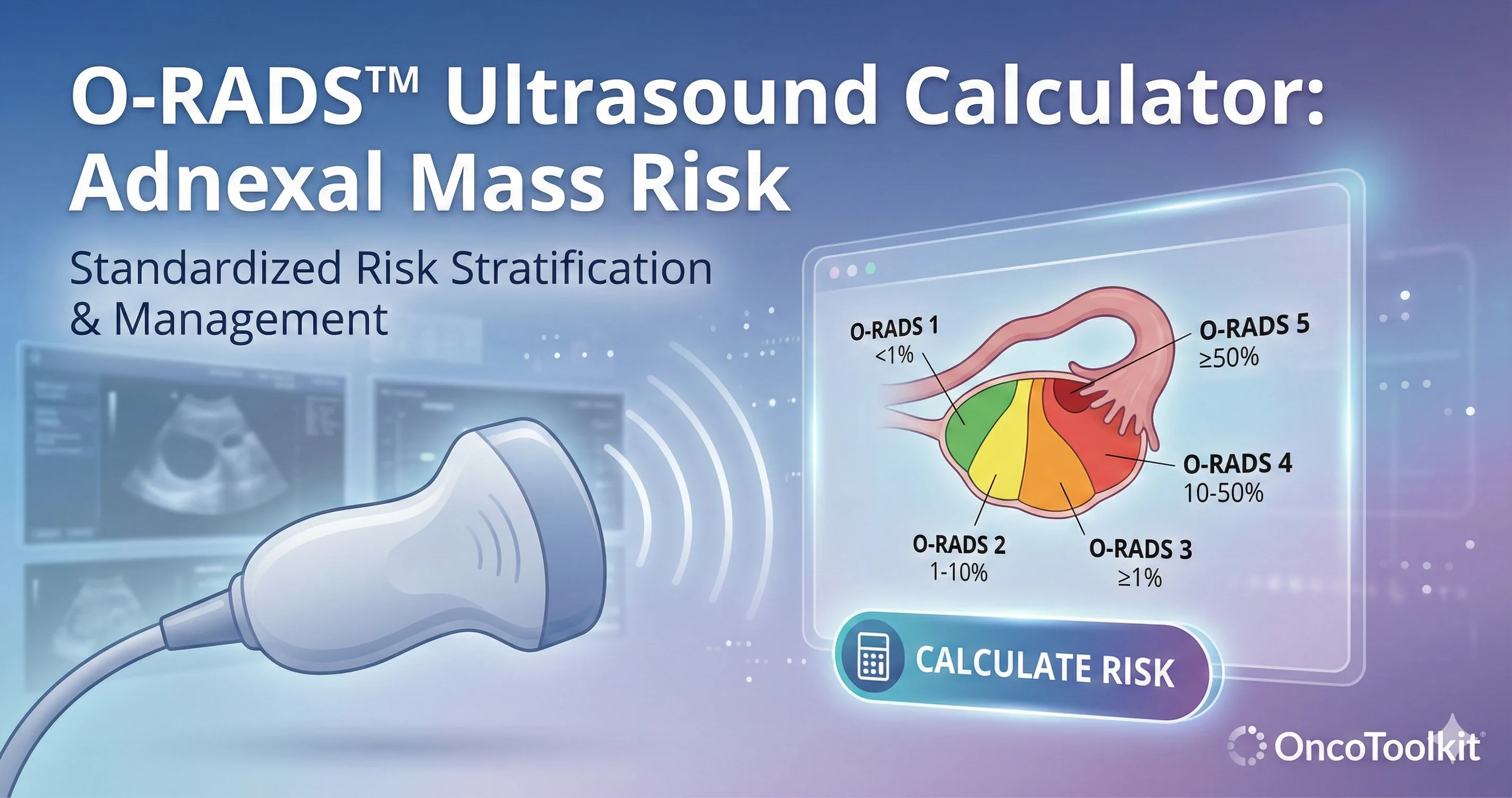
O-RADS™ (Ultrasound) calculator: fast, standardized adnexal mass risk stratification
Learn how to move from sonographic descriptors to clear risk categories and management plans using the official ACR O-RADS US framework.
Quick Navigation
- 1. Introduction to O-RADS™ Ultrasound
- 2. Understanding the O-RADS™ US Risk System
- 3. The Clinical Importance of O-RADS™ in Practice
- 4. Evidence, Risk Categories, and Algorithmic Logic
- 5. User Guide: How the OncoToolkit Calculator Works
- 6. Supporting Care, Education, and Clinical Research
- 7. Clinical FAQ: Common Questions and Pitfalls
- 8. References
1. Introduction to O-RADS™ Ultrasound
Ultrasound assessment of adnexal masses is central to gynecologic practice but often challenging, especially when distinguishing physiologic cysts from lesions that warrant oncologic referral. Multiple sonographic descriptors, menopausal status, and varying malignancy risk estimates must be integrated quickly, often under time pressure in clinic, the ED, or MDT meetings. At OncoToolkit, we have built an O-RADS™ (Ultrasound) calculator that implements the official ACR O-RADS US risk stratification and management system, allowing gynecologists and gynecologic oncologists to move from key ultrasound features to a clear risk category and suggested management in a few taps. [1], [2], [3], [4], [5]
The calculator is applicable to both premenopausal and postmenopausal patients, with inputs designed to mirror how the ACR lexicon describes adnexal lesions in routine practice. Color‑coded visual output, approximate malignancy risk, and plain‑language management prompts help reduce cognitive load while keeping you aligned with guideline-based care. [2], [6], [3], [4]
2. Understanding the O-RADS™ US Risk System
O-RADS™ US is a structured reporting and risk stratification system developed by the American College of Radiology (ACR) for ultrasound assessment of ovarian and adnexal masses. It categorizes lesions from O-RADS 0–5 based on morphology, lesion size, and vascularity, with each category corresponding to an estimated risk of malignancy and a linked management strategy. [6], [3], [4]
Core Features of O-RADS US:
- Uses a standardized lexicon for morphology (e.g., unilocular cyst, multilocular cyst, solid component). [4], [5]
- Incorporates lesion dimensions and the so‑called “3 mm rule” for papillary or solid components. [5], [4]
- Integrates Doppler findings using the IOTA color score (1–4) as a surrogate for vascularity. [7], [4]
The system has been iteratively refined (from v1 to v2022) and is now widely taught in radiology and gynecology, with multiple validation and comparative studies showing good diagnostic performance for differentiating benign from malignant adnexal masses. [8], [9], [1]
3. The Clinical Importance of O-RADS™ in Practice
For gynecologists and gyn oncologists, adnexal mass workup has direct implications for surgical planning, referral, and counseling about fertility and staging procedures. O-RADS US provides a common language and risk framework so that sonographers, radiologists, and surgeons can communicate clearly about the likelihood of malignancy and the appropriate next step. [10], [11], [3], [4]
Without a digital implementation, applying O-RADS at the point of care can be cumbersome:
- • Morphologic categories and subcategories are numerous and nuanced. [2], [5]
- • Risk percentage ranges and thresholds for surgery or referral can be difficult to recall. [3], [2]
- • Clinicians may juggle other models (e.g., IOTA ADNEX) alongside O-RADS. [11]
On our platform, the O-RADS™ (Ultrasound) calculator translates this complexity into a short, responsive form.
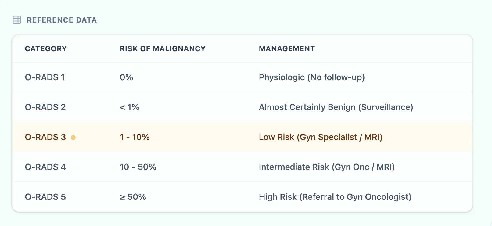
Figure 2. Reference table linking risk categories to suggested management.
4. Evidence, Risk Categories, and Algorithmic Logic
4.1 O-RADS™ Categories and Malignancy Risk
4.2 Underlying Logic: The Decision Tree Approach
Unlike regression-based models, O-RADS US functions as an algorithmic decision tree determined by: [4], [5]
- Menopausal status.
- Dominant morphology pattern.
- Maximum lesion diameter.
- Vascularity (IOTA color score 1–4). [7], [4]
- Modifiers (e.g., acoustic shadows). [9], [11]
4.3 Validation Data and Clinical Limitations
- High sensitivity for malignant lesions.
- Observed malignancy frequencies matching guideline-defined ranges.
- Moderate agreement with alternative models like IOTA ADNEX. [11]
Clinical Note: O-RADS US is for adult patients at average risk without acute emergent symptoms. Local protocols should take precedence in emergent cases. [13], [10]
5. User Guide: How the OncoToolkit Calculator Works
5.1 Input Parameters for Ultrasound Reporting
The calculator uses five main inputs:
- Menopausal Status | 2. Dominant Morphology | 3. Max Diameter (cm) | 4. Vascularity | 5. Acoustic Shadows
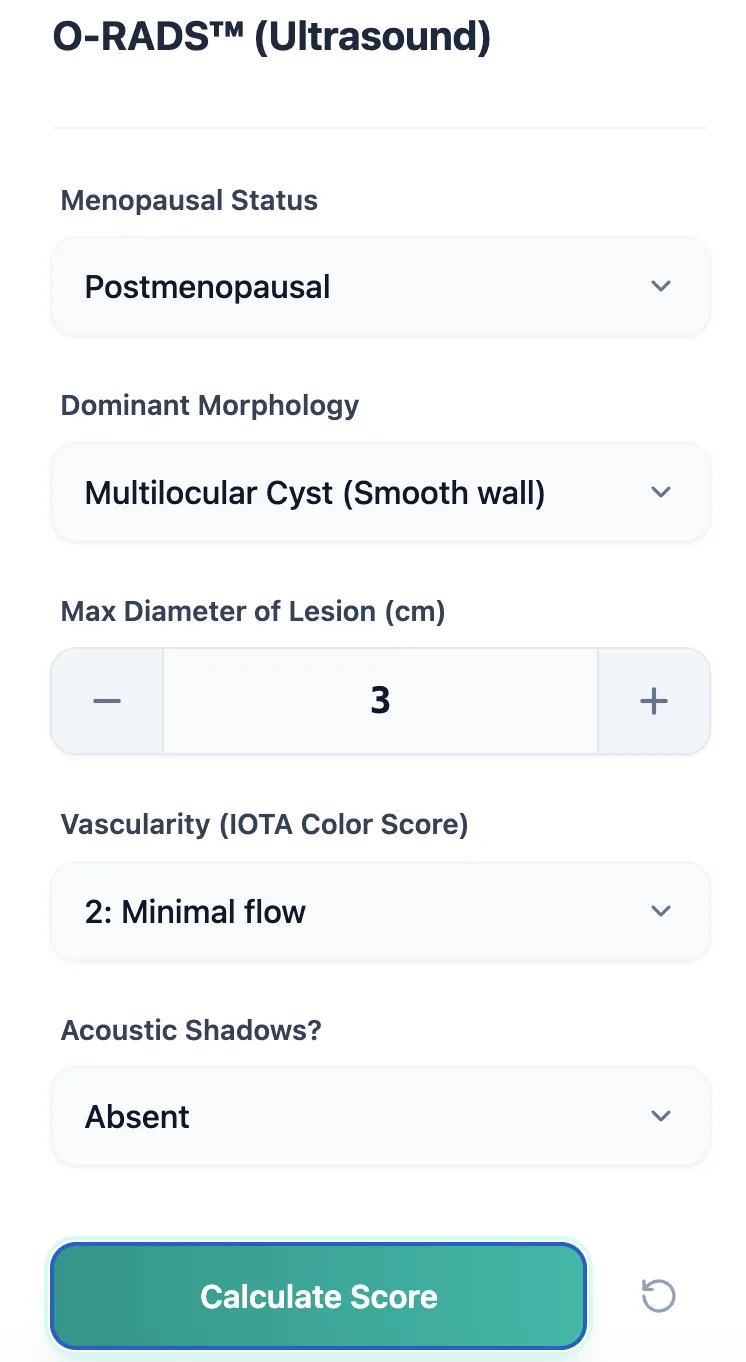
Figure 3. The input form matches the O-RADS US lexicon used in routine reporting.
5.2 Interpreting Category, Risk, and Management Outputs
- O-RADS category (0–5).
- Risk profile band.
- Management suggestion (Surveillance, MRI, or Oncology Referral). [10], [3], [6]
- Color‑coded bar for visual reinforcement.
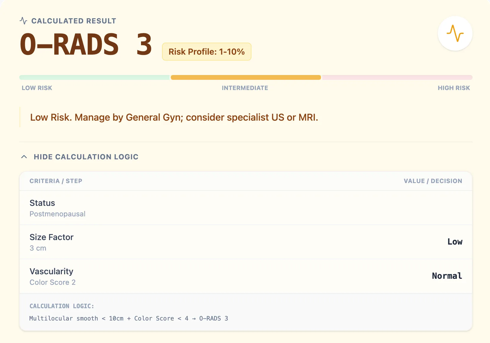
Figure 4. Example result showing low-risk category and management logic.
6. Supporting Care, Education, and Clinical Research
Routine Support
Standardizing assessment and facilitating MDT communication. [11], [3]
Trainee Education
Using the expanded logic view to teach branching rules. [2], [5]
Research & QI
Exporting categories to connect with pathology outcomes. [9], [11]
7. Clinical FAQ: Common Questions and Pitfalls
When should you not rely solely on this tool?
Not for hemodynamically unstable patients, acute pain, or very high genetic risk (BRCA carriers). [13], [10]
How does it compare with IOTA ADNEX?
O-RADS gives discrete categories/management; ADNEX gives granular percentages. Both perform well and can be used in combination. [9], [11]
What is the most common mistake?
Misclassifying small papillary projections or omitting lesion size thresholds. [5], [2]
Ready to Simplify Adnexal Risk Stratification?
Quickly convert ultrasound findings into actionable ACR-aligned management plans.
Try the O-RADS™ Calculator Now
Free to use. No registration required.
8. References
- JAMA Network Open - O-RADS Performance Source
- RadiologyKey - O-RADS US v2022 Source
- VolusonClub - O-RADS Guidelines Source
- PubMed - O-RADS US Lexicon Source
- AJR - Lexicon and Category Review Source
- Radiology UW - Assessment Categories Source
- PMC - IOTA Color Score Source
- AJR - Validation Study Source
- PMC - Meta-analysis of O-RADS Source
- Cancer Care Ontario - Standardized Reporting Source
- PMC - Comparison with IOTA ADNEX Source
- RadAtHand - Calculator Logic Source
- ACR Support - Applicability Criteria Source